Anterior Cruciate Ligament (ACL) Function & Anatomy
Anterior Cruciate Ligament (ACL) Function & Anatomy
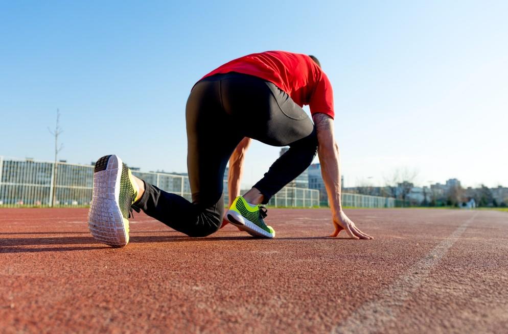
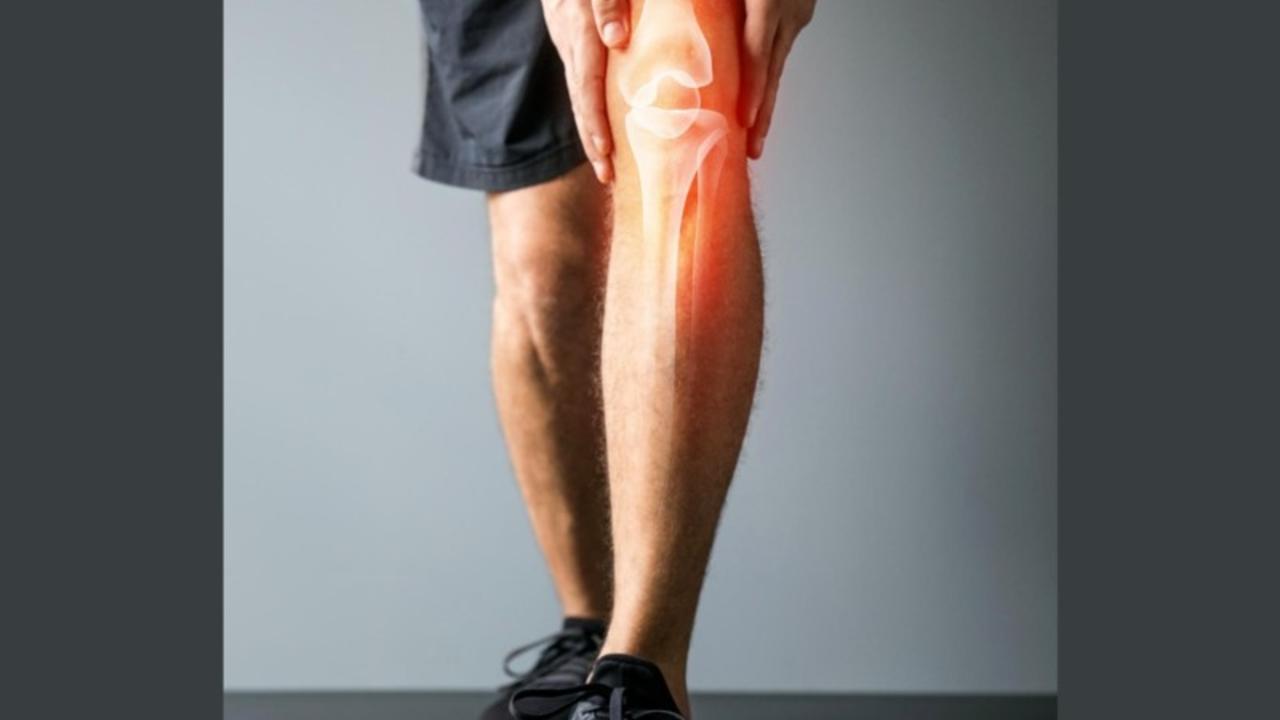
The Anterior Cruciate Ligament (ACL) is an essential structure within your knee. The ACL plays a crucial role in stabilizing your knee by limiting rotation and avoiding overextension of the Tibia (shin bone). However, in many high-impact sports (Basketball, Football, Soccer, etc.), a lot of pressure is put on the ACL, making it one of the most often injured ligaments in sports. The problem here is that the ACL does not heal by itself and, therefore, usually requires surgery to restore the injured knee's stability.

Knee Anatomy
The picture depicts the knee's main anatomical structures (here looking at the right knee from the front). Moving from top to bottom, we start with the Femur. The Femur is your thigh bone and the strongest bone of the human body. On top of the Femur are the Quadriceps muscles. As their name suggests, the Quadriceps consists of 4 major muscles (Rectus femoris, Vastus intermedius, Vastus lateralis & Vastus Medialis). Besides its use to flex the hip (lifting your knee upwards in front of you when standing), the Quadriceps allow you to extend your leg by straightening your knee. Throughout this movement, the Quadriceps contract, pulling on the Quadriceps tendon. The Quadriceps tendon attaches to your Patella (Kneecap). The energy is then transferred over the Patellar tendon, which connects the Kneecap with your Tibia (Shinbone). On the outside of your knee, the Lateral collateral ligament (LCL) connecting your Femur and the Fibula. The Fibula is the small bone in your lower leg. Your Medial collateral ligament (MCL) connects the Femur to the Tibia on the inside of your knee.
Having worked ourselves around the knee, let us take a look inside. Here you can find the Meniscus, the Anterior cruciate ligament (ACL) and the Posterior cruciate ligament (PCL). You have two Menisci (lateral and medial) consisting of cartilage and acting as a shock absorber between your Femur and Tibia. The ACL and PCL also connect the Femur and the Tibia. The PCL runs from the medial femoral condyle (inside part of the Articular cartilage or smooth part at the end of the Femur at the joint) to the posterior of the Tibial plateau (top and back of Tibia bone). Finally, the ACL runs from the posterior (back) of the Lateral (femoral) condyle to the front of the Tibia, crossing the PCL. This is why these two ligaments are called the cruciate (cross-shaped) ligaments.
ACL Function
The ACL stabilizes your knee. When you walk, momentum will move your Tibia forward, which is called anterior force. Firstly, to ensure your knee stays aligned, the ACL restrains about 85% of this anterior force. Secondly, when walking or running, both the Femur and Tibia rotate in- and externally. The ACL also plays a vital role in restricting these rotational forces.
ACL Injury
An ACL injury, therefore, affects the stability of your knee. This missing stability can ultimately lead to structural damages within the knee joint. The ACL ruptures or is injured when the forces exerted on it are too great (usually rotational force) as through.
Summary
The ACL plays a vital role in knee stability. If injured or ruptured, it is instrumental to get it fixed through surgery or extensive physical therapy. Otherwise, there can be long-term damages. It is possible to compensate for a ruptured ACL through strength training and pronounced musculature; however, this will certainly not avoid long-term damage. In either case, it is crucial to knee stability to strengthen your lower extremities and core.
Author

Chris
Author
Hi! My name is Chris, I am 25 years old and I created Athletic-Recovery.com, because I want to help people recover from sports injuries. Personally, I ruptured my ACL in 2017 and my Achilles Tendon in 2020. Both injuries were great set backs, but motivated me to create something where people going through the same struggles all around the world can share their experiences and knowledge to help each other get through their personal injury. #recovertogether